Beta bursts during continuous movements accompany the velocity decrement in Parkinson's disease patients.
Bursts of pathological brain waves occur as patients with Parkinson’s make voluntary movements. These bursts may contribute to the characteristic slowing of movements that occurs in this condition.
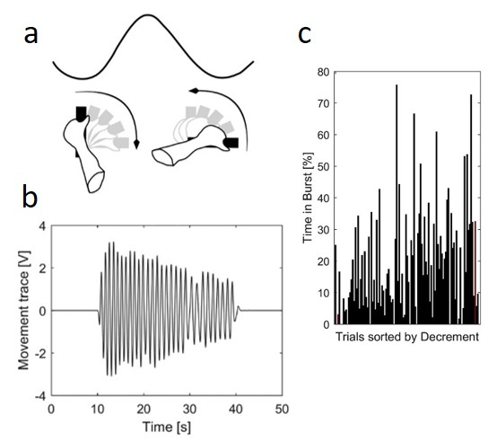
Bradykinesia is reported to correlate with subthalamic beta power (13-35 Hz) recorded at rest in Parkinson's disease (PD). Pilot studies suggest adaptive deep brain stimulation triggered by amplitude threshold crossings of beta activity defined at rest is effective. This is puzzling, given that beta is suppressed during repetitive movements when bradykinesia becomes apparent. Recently, increased beta power in PD has been linked to beta bursts. Here we investigate whether beta bursts also occur during repetitive movements and relate to progressive decrement in movement velocity. Therefore, subthalamic local field potentials were recorded in 12 PD patients off medication while performing 30s blocks of rotatory movements alternating with rest periods. Bursts were defined separately for the low (13-20 Hz) and high (20-35 Hz) beta band using thresholds defined at rest. As expected, velocity significantly decreased within movement blocks. Despite the sustained suppression of both beta sub-bands, bursts could still be detected during movement. Beta bursts were reduced in amplitude, duration and rate during movement with beta rate correlating best with beta power. A mixed-effects linear model revealed that percentage time spent in beta bursts predicted velocity decreases better than averaged power. This correlation was specific for the low beta band. Our results link beta bursts during movement to bradykinesia. This helps explain how beta activity may contribute to bradykinetic movement decrement even though mean beta power is reduced during movement. Moreover, our findings help explain the effectiveness of adaptive DBS triggered off beta bursts, even though these may be defined with respect to beta levels at rest.
2020. J. Neurosci., 40(7):1571-1580.