Waveform changes with the evolution of beta bursts in the human subthalamic nucleus.
Bursts of brain nerve cell activity at a particular frequency have been linked to movement difficulties in people with Parkinson’s. Here, we used sophisticated analysis techniques to show how these bursts appear and disappear in a deep brain area important for movement. This affords us clues as to what brain processes might be involved in the termination of pathological bursts.
Objective: Phasic bursts of beta band synchronisation have been linked to motor impairment in Parkinson’s disease (PD). However, little is known about what terminates bursts.
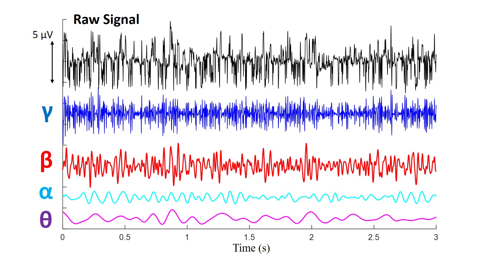
Methods: We used the Hilbert–Huang transform to investigate beta bursts in the local field potential recorded from the subthalamic nucleus in nine patients with PD on and off levodopa.
Results: The sharpness of the beta waveform extrema fell as burst amplitude dropped. Conversely, an index of phase slips between waveform extrema, and the power of concurrent theta activity increased as burst amplitude fell. Theta activity was also increased on levodopa when beta bursts were attenuated. These phenomena were associated with reduction in coupling between beta phase and high gamma activity amplitude. We discuss how these findings may suggest that beta burst termination is associated with relative desynchronization of the beta drive, increase in competing theta activity and increased phase slips in the beta activity.
Conclusions: We characterise the dynamical nature of beta bursts, thereby permitting inferences about underlying activities and, in particular, about why bursts terminate.
Significance: Understanding the dynamical nature of beta bursts may help point to interventions that can cause their termination and potentially treat motor impairment in PD.