Beta Burst Characteristics and Coupling within the Sensorimotor Cortical-Subthalamic Nucleus Circuit Dynamically Relate to Bradykinesia in Parkinson's Disease.
Yao, Sharma et al. studied prolonged brain activity recordings from patients with Parkinson’s disease (PD) implanted with sensing enabled deep brain stimulation (DBS) devices. By relating this brain activity to concurrent measurements of symptom severity, obtained via wristwatch worn devices, they identify brain activity patterns related to movement slowing in PD. These findings could help to personalise and improve the effectiveness of DBS for PD.
Background
Bursts of exaggerated subthalamic nucleus (STN) beta activity are believed to contribute to clinical impairments in Parkinson's disease (PD). No previous studies have explored burst characteristics and coupling across the sensorimotor cortical-STN circuit and determined their relationship to dynamic measurements of bradykinesia.
Objective
We sought to (1) establish the characteristics of sensorimotor cortical and STN bursts during naturalistic behaviors, (2) determine the predictability of STN bursts from motor cortical recordings, and (3) relate burst features to continuous measurements of bradykinesia using wearable sensors.
Methods
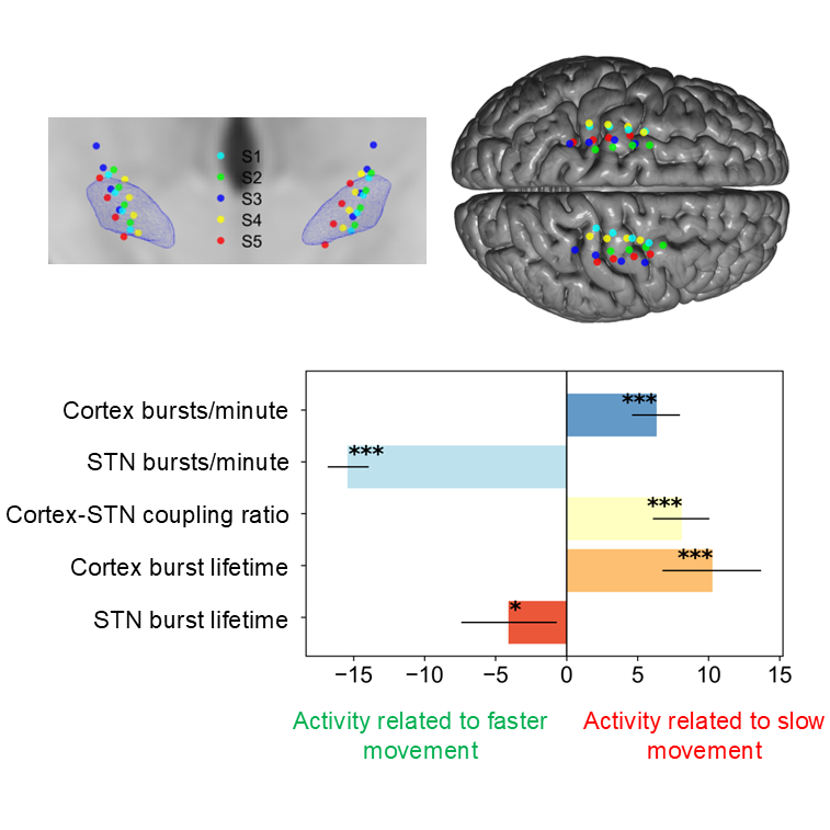
We analyzed 1046 h of wirelessly streamed bilateral sensorimotor cortical and STN recordings from 5 PD patients with concurrent measurements of bradykinesia.
Results
STN bursts were longer than cortical bursts and had shorter inter-burst intervals. Long bursts (>200 ms) in both structures displayed temporal overlap (>30%), with cortical bursts tending to lead STN burst onset by 8 ms. Worsening bradykinesia was linked to increased cortical burst rates and durations, whereas STN burst properties had the opposite effect.
Conclusion
Cortical beta bursts tend to precede STN beta bursts with short delays and their occurrence relates to worsening bradykinesia in naturalistic settings.
2025. Neurobiol Dis, 207:106858.
2022. NPJ Parkinsons Dis, 8(1):88.
2025. Mov Disord, 40(3):456-467.
2023. Brain, 146(12):5015-5030.