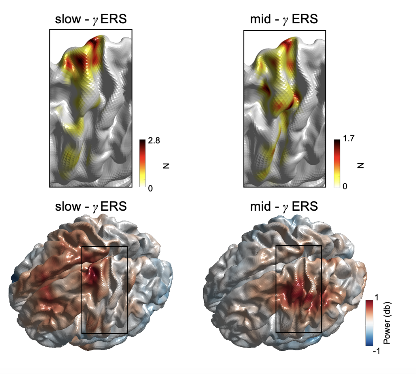
Human motor cortical gamma activity relates to GABAergic intracortical inhibition and motor learning.
Here, Zich and colleagues studied a certain type of brain waves called gamma waves (frequency >30 Hz) during movement. Using brain scans, stimulation, and movement tasks, they identified two distinct gamma patterns: slow-gamma (30-60 Hz) linked to inhibitory brain chemicals, and mid-gamma (60-90 Hz) associated with motor learning. These findings help explain how gamma brain waves support human movement and skill acquisition.
Gamma activity (γ, >30 Hz) is universally demonstrated across brain regions and species. However, the physiological basis and functional role of γ sub-bands (slow-γ, mid-γ, fast-γ) have been predominantly studied in rodent hippocampus; γ activity in the human neocortex is much less well understood. We use electrophysiology, non-invasive brain stimulation, and several motor tasks to examine the properties of sensorimotor γ activity sub-bands and their relationship with both local GABAergic activity and motor learning. Data from three experimental studies are presented. Experiment 1 (N = 33) comprises magnetoencephalography (MEG), transcranial magnetic stimulation (TMS), and a motor learning paradigm; experiment 2 (N = 19) uses MEG and motor learning; and experiment 3 (N = 18) uses EEG and TMS. We characterised two distinct γ sub-bands (slow-γ, mid-γ) which show a movement-related increase in activity during unilateral index finger movements and are characterised by distinct temporal-spectral-spatial profiles. Bayesian correlation analysis revealed strong evidence for a positive relationship between slow-γ (~30-60 Hz) peak frequency and GABAergic intracortical inhibition (as assessed using the TMS-metric short interval intracortical inhibition). There was also moderate evidence for a relationship between the power of the movement-related mid-γ activity (60-90 Hz) and motor learning. These relationships were neurochemical and frequency specific. These data provide new insights into the neurophysiological basis and functional roles of γ activity in human M1 and allow the development of a new theoretical framework for γ activity in the human neocortex.

2025. Imaging Neurosci (Camb), 3.
2021. eLife, 10:e67355
2022. Exp Neurol, 351:113999.
