Magill Group
Our overall goal is to provide detailed explanations of how brain circuit organisation supports normal and impaired behaviours. Focusing on a brain region called the basal ganglia, we monitor and manipulate different types of nerve cell to provide new insights into how their host networks operate. In taking advantage of the new understanding gained, we use specialised nerve cell types as entry points for novel therapeutic interventions that are designed to correct the brain circuit disorganisation and behavioural difficulties that arise in disease.
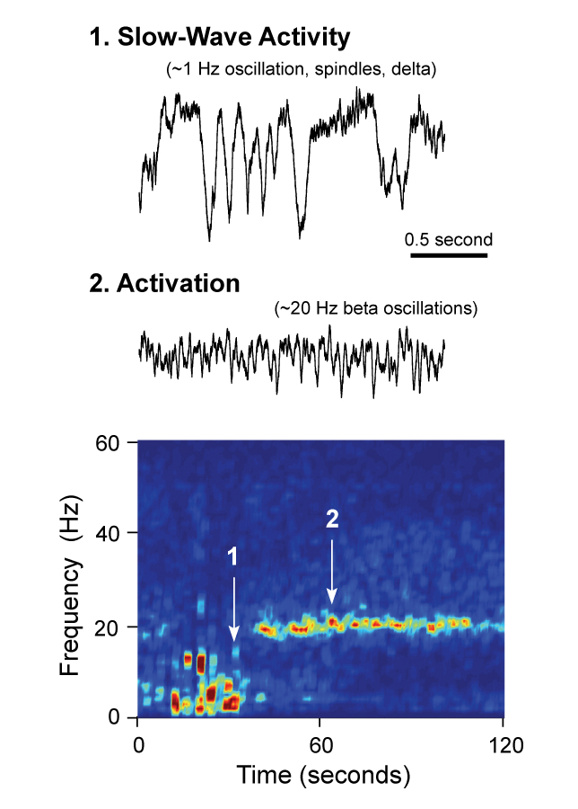
We recognise that the burden of disease is not borne evenly across all cell types in the brain. It is thus imperative that the design of new strategies for treating disease symptoms is tempered by a mature knowledge of how different cell types fulfil their specialised roles to govern behaviour. The overarching goal of our Programme is to fill this knowledge gap by delivering high-resolution readouts and mechanistic explanations of brain 'motor circuit' organization in the context of normal behaviours as well as impaired behaviours. Focusing on basal ganglia and thalamocortical circuits, we harness cutting-edge technologies for identifying, monitoring, accessing and manipulating neurons in vivo to provide fundamental new insights into the specific cellular substrates of the neuronal network dynamics therein. We place special emphasis on defining how the interactions and activities of identified cell types in these brain circuits vary according to the temporal profile of dopamine release and movement. As a key corollary of this, we define how a paucity of dopamine release, as occurs in Parkinson’s disease and its animal models, impacts on the neuronal encoding of behaviour in these motor circuits. In capitalising on the new level of understanding of the dynamics of identified neurons that is gained here, we also endeavour to exploit specified cell types and other circuit elements as novel points of entry for spatiotemporally-patterned interventions designed to not only dissect circuit function but also to correct circuit dysfunction and related behavioural deficits in Parkinsonism and other disorders of movement and memory.
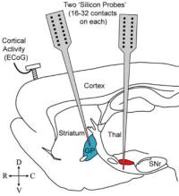
We couple novel and advanced analytical techniques with experimental interventions that probe causal interactions between specified circuit elements with high spatiotemporal precision. Our experiments centre on the use of wild type and genetically-altered rodents with intact or comprised midbrain dopaminergic systems, the readouts from which straddle multiple levels of function including molecular/genetic, structural, electrophysiological, neurochemical and behavioural.
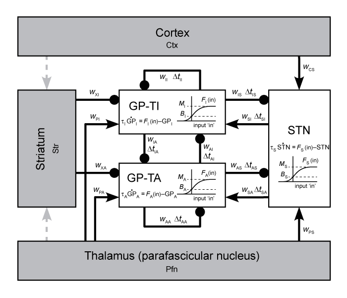
- Mechanisms underlying neuronal network activity in basal ganglia-thalamocortical circuits.
- Cell-type-selective encoding of behaviour in basal ganglia-thalamocortical circuits.
- Experimental models of movement/memory disorders involving basal ganglia-thalamocortical circuits.
- Generation, dissemination and impact of aberrant neuronal activity in the Parkinsonian brain.
- Cell-type-selective interventions for symptom relief in disease.
Our research is designed to provide significant advances in the understanding of how specialised cell types in the basal ganglia work together with neurons in their partner brain circuits to control behaviour, for better or worse. We recognise that new understanding is critically important for building a stronger foundation from which to develop new therapeutic interventions in disease. We thus strive to progress from delivering new mechanistic insights, through generation of firm rationale to proof-of-concept studies that can be taken forward to inform and advance the future development of improved, personalised therapies.
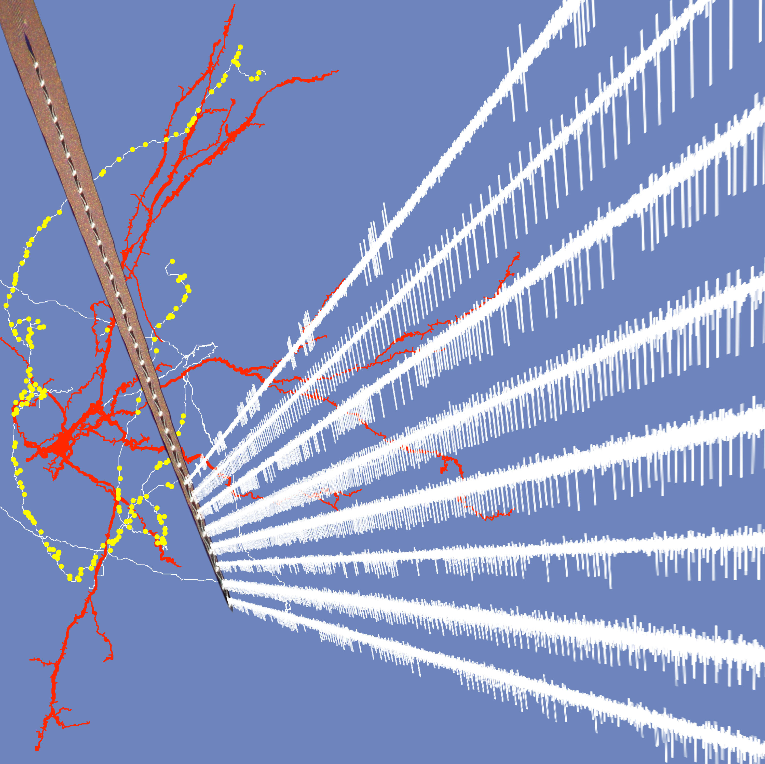
- Electrophysiology (in vivo and in vitro)
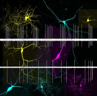
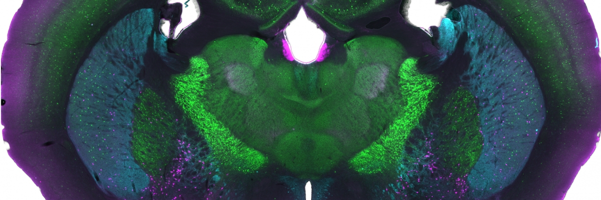
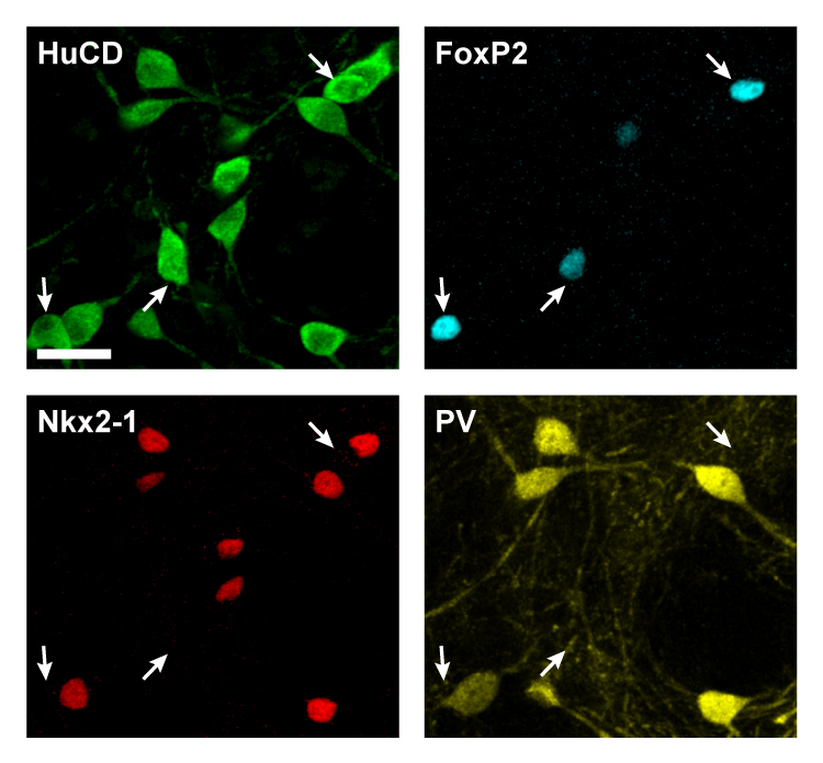
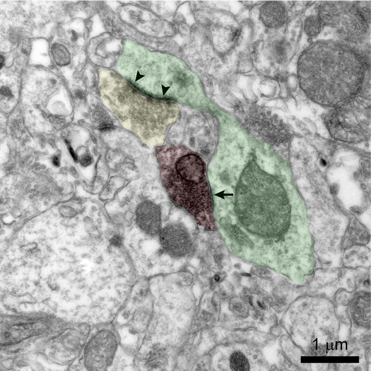
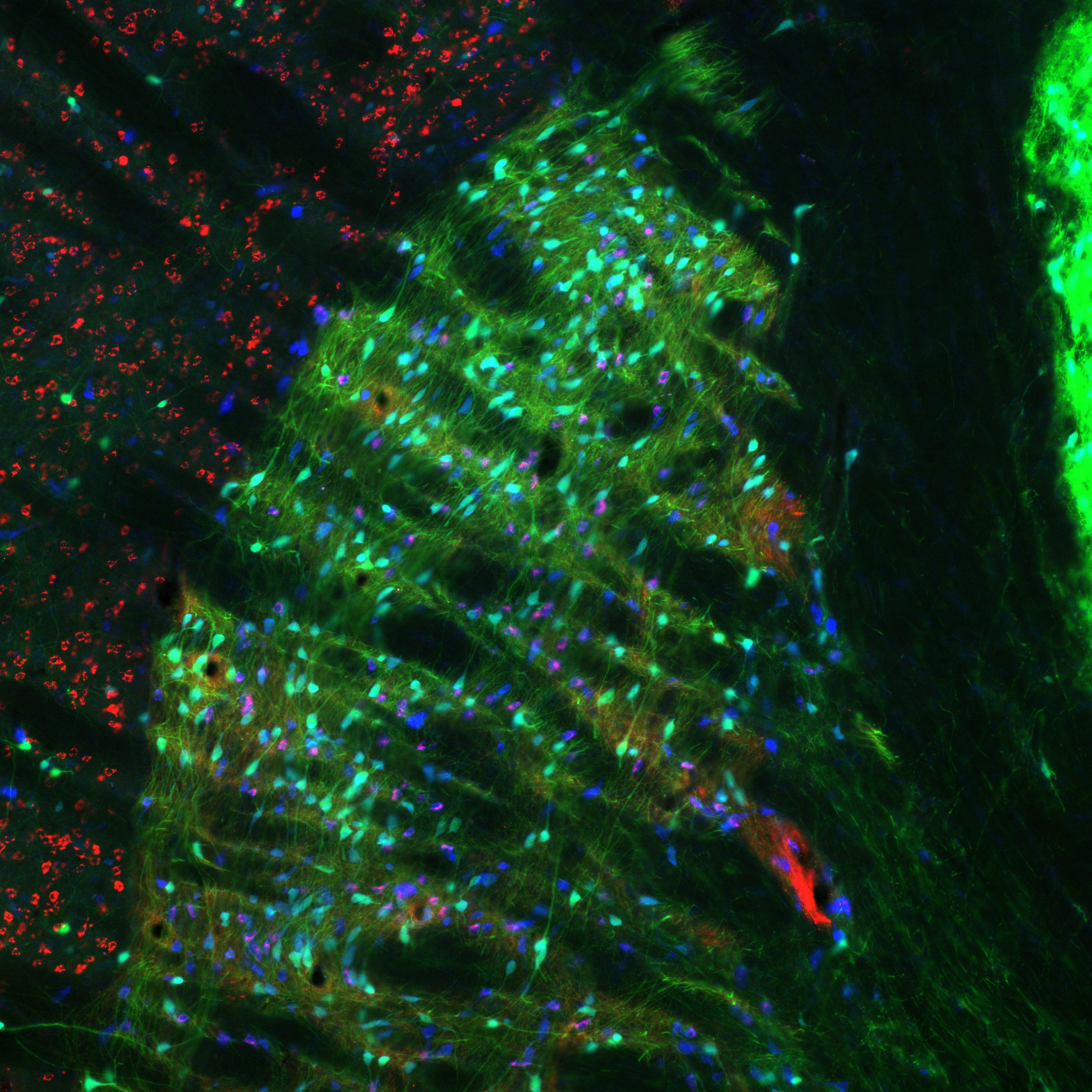
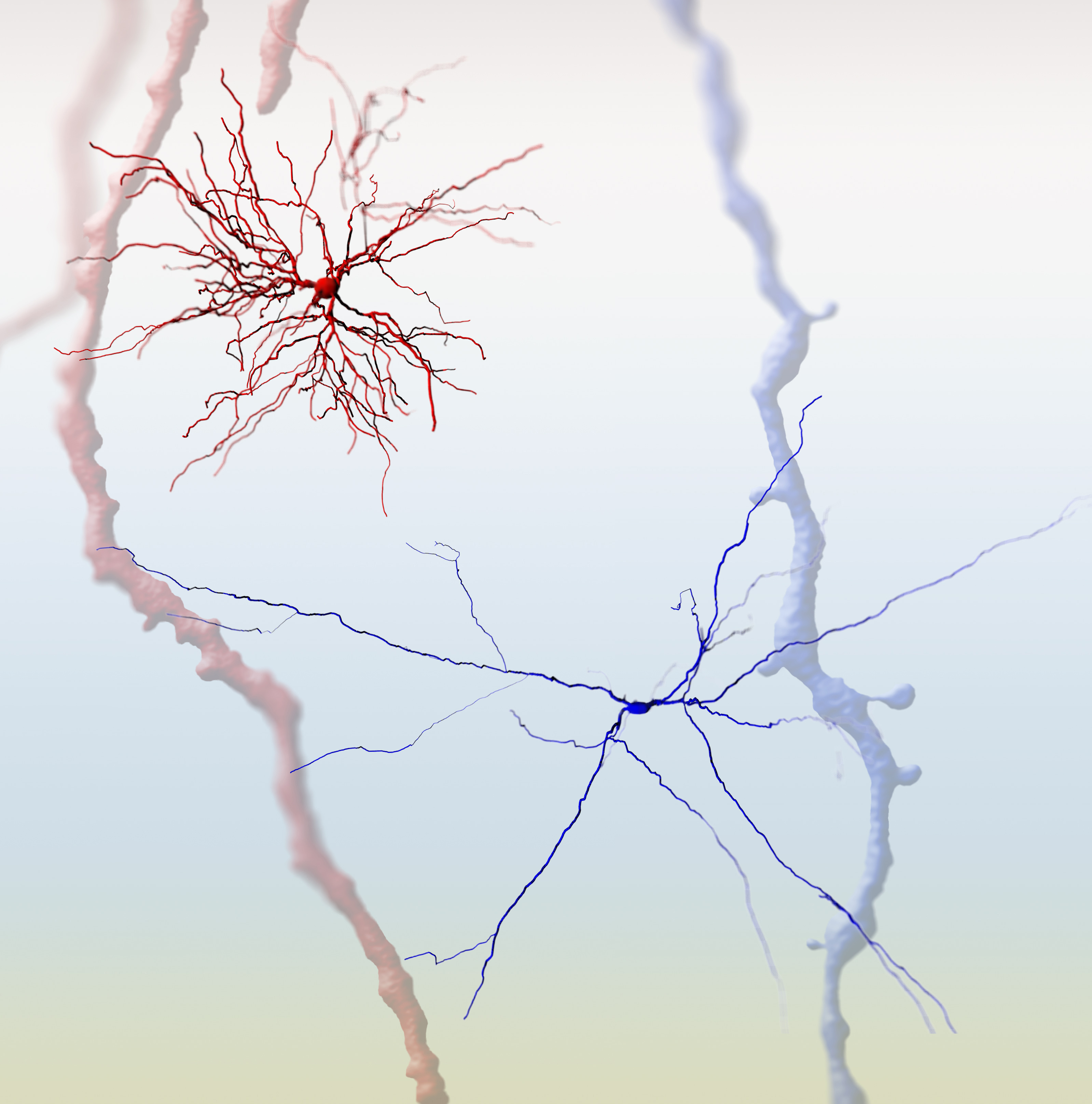
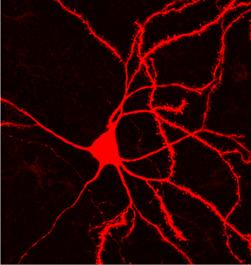
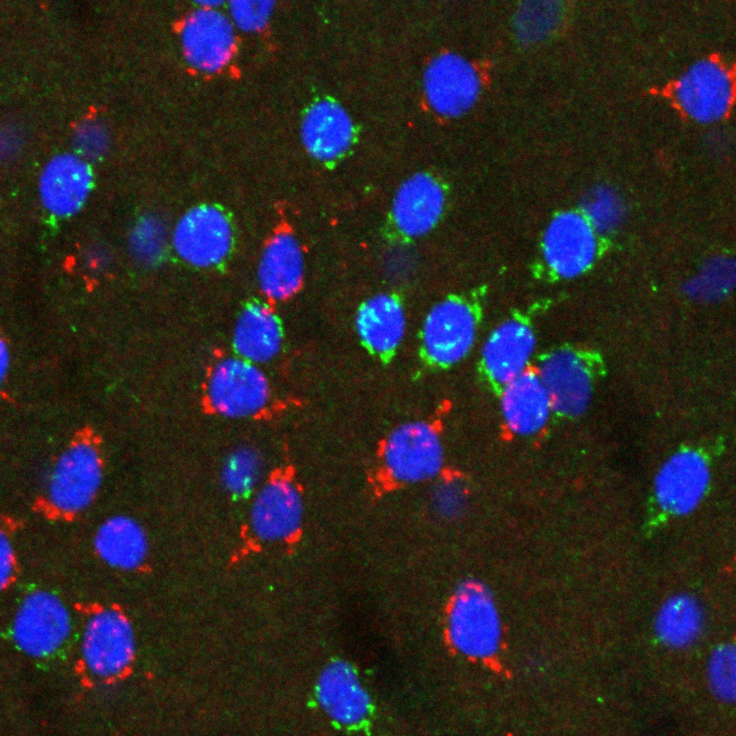
- Light and electron microscopy
- Genetics-based approaches for cell monitoring and manipulation
- Quantification of voluntary behaviours
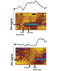
- Fibre photometry and fast-scan cyclic voltammetry in vivo
We are committed to fostering an inclusive work environment that celebrates diversity and promotes equal opportunity within our group and the wider MRC BNDU.


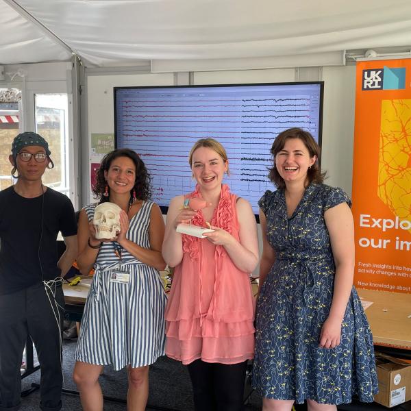
The enthusiastic BNDU team of scientists. (L-R) Shenghong, Chiara, Rosie and Ioana.
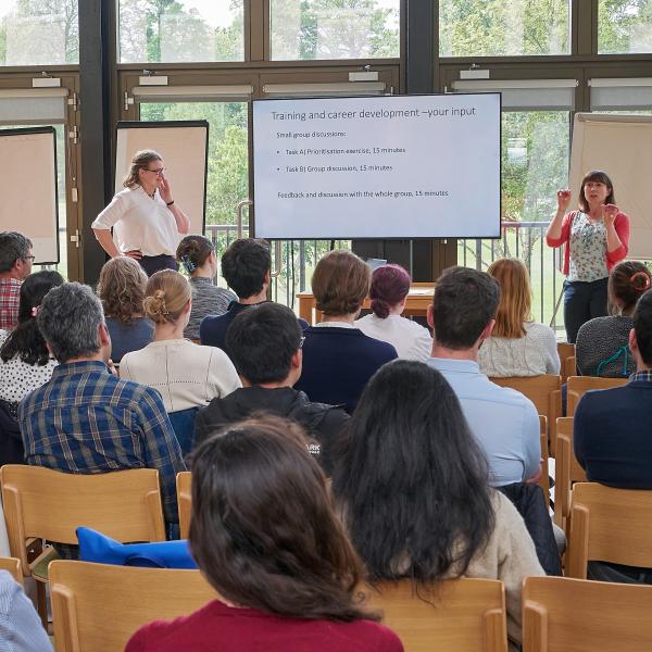
Mary Muers (far right) moderates an interactive session about training and career development opportunities.
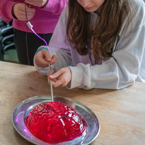
A visiting school pupil tries their hand at implanting a dummy stimulation electrode in a jelly brain!
Recent Preprints
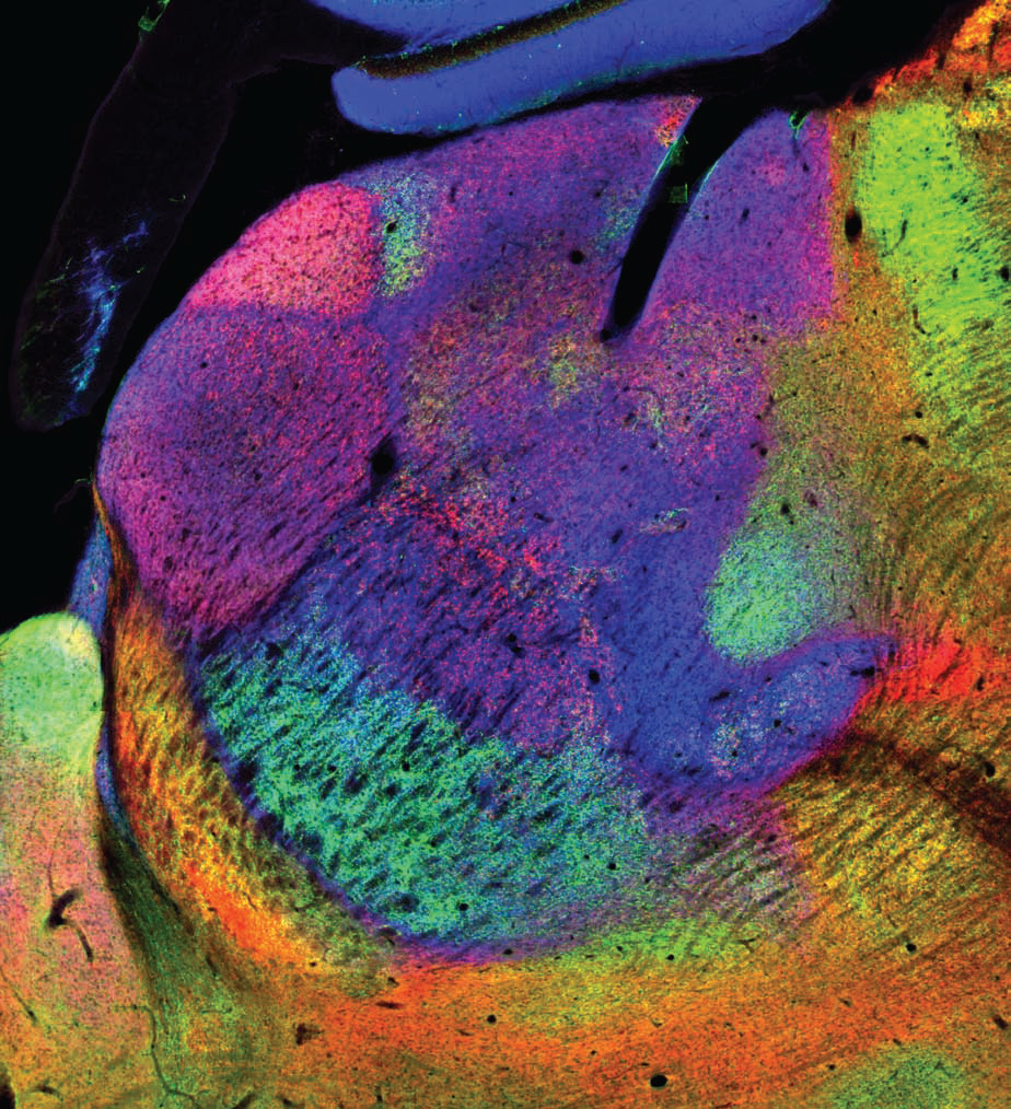
Like other Groups at the MRC BNDU, we are committed to best practice in open research. We have created and curated a range of primary data, metadata and related resources that can be readily downloaded by external users from the MRC BNDU's Data Sharing Platform. We are part of the team that created CHAMBER, a novel chemoarchitectonic atlas of mouse thalamus and other brain regions. We highlight below just a few examples of the other datasets and resources we have shared for the benefit of our stakeholders.