Local field potential activity dynamics in response to deep brain stimulation of the subthalamic nucleus in Parkinson's disease.
One of the key goals of the MRC Brain Network Dynamics Unit is to develop better controlled deep brain stimulation for the treatment of brain disorders. We are particularly interested in using brain signals to control when and how much stimulation is delivered. Here, we study the characteristics of deep brain signals to help understand how they may be used to control stimulation.
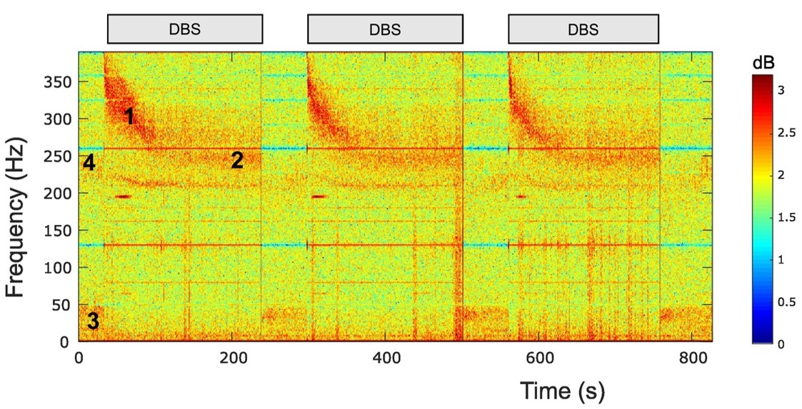
Local field potentials (LFPs) may afford insight into the mechanisms of action of deep brain stimulation (DBS) and potential feedback signals for adaptive DBS. In Parkinson's disease (PD) DBS of the subthalamic nucleus (STN) suppresses spontaneous activity in the beta band and drives evoked resonant neural activity (ERNA). Here, we investigate how STN LFP activities change over time following the onset and offset of DBS. To this end we recorded LFPs from the STN in 14 PD patients during long (mean: 181.2 s) and short (14.2 s) blocks of continuous stimulation at 130 Hz. LFP activities were evaluated in the temporal and spectral domains. During long stimulation blocks, the frequency and amplitude of the ERNA decreased before reaching a steady state after ~70 s. Maximal ERNA amplitudes diminished over repeated stimulation blocks. Upon DBS cessation, the ERNA was revealed as an under-damped oscillation, and was more marked and lasted longer after short duration stimulation blocks. In contrast, activity in the beta band suppressed within 0.5 s of continuous DBS onset and drifted less over time. Spontaneous activity was also suppressed in the low gamma band, suggesting that the effects of high frequency stimulation on spontaneous oscillations may not be selective for pathological beta activity. High frequency oscillations were present in only six STN recordings before stimulation onset and their frequency was depressed by stimulation. The different dynamics of the ERNA and beta activity with stimulation imply different DBS mechanisms and may impact how these activities may be used in adaptive feedback.
2023. Neuromodulation, 26(2):320-332.
2023. Mov Disord, 38(5):818-830.