GABAergic Medial Septal Neurons with Low-Rhythmic Firing Innervating the Dentate Gyrus and Hippocampal Area CA3.
Cells in part of the brain called the medial septum can be classified into two types by how rhythmically they fire action potentials. In this study in mice, the low rhythmic cells were compared to the high, both in terms of where their outputs are in the brain, and timing of their firing. The outputs in two areas of the hippocampus, and their firing patterns, suggest that they could be involved in memory discrimination.
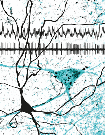
The medial septum implements cortical theta oscillations, a 5-12 Hz rhythm associated with locomotion and paradoxical sleep reflecting synchronization of neuronal assemblies such as place cell sequence coding. Highly rhythmic burst-firing parvalbumin-positive GABAergic medial septal neurons are strongly coupled to theta oscillations and target cortical GABAergic interneurons, contributing to coordination within one or several cortical regions. However, a large population of medial septal neurons of unidentified neurotransmitter phenotype and with unknown axonal target areas fire with a low degree of rhythmicity. We investigated whether low-rhythmic-firing neurons (LRNs) innervated similar or different cortical regions to high-rhythmic-firing neurons (HRNs) and assessed their temporal dynamics in awake male mice. The majority of LRNs were GABAergic and parvalbumin-immunonegative, some expressing calbindin; they innervated interneurons mostly in the dentate gyrus (DG) and CA3. Individual LRNs showed several distinct firing patterns during immobility and locomotion, forming a parallel inhibitory stream for the modulation of cortical interneurons. Despite their fluctuating firing rates, the preferred firing phase of LRNs during theta oscillations matched the highest firing probability phase of principal cells in the DG and CA3. In addition, as a population, LRNs were markedly suppressed during hippocampal sharp-wave ripples, had a low burst incidence, and several of them did not fire on all theta cycles. Therefore, CA3 receives GABAergic input from both HRNs and LRNs, but the DG receives mainly LRN input. We propose that distinct GABAergic LRNs contribute to changing the excitability of the DG and CA3 during memory discrimination via transient disinhibition of principal cells.
2024. Neuron, 112(22):3768-3781.e8.
