Hippocampal Theta Input to the Amygdala Shapes Feedforward Inhibition to Gate Heterosynaptic Plasticity.

Bazelot M
Bocchio M
Kasugai Y
Fischer D
Dodson PD
Ferraguti F
Capogna M

The amygdala and the hippocampus are two brain structures involved in emotional behaviour. Disrupted hippocampus-amygdala interplay appears to occur in anxiety disorders, but the mechanisms of communication between these two brain areas are largely unknown. In this work, we have addressed this issue and discovered that inhibitory nerve cells that use the chemical GABA coordinate such interplay.

Video Abstract
Scientific Abstract

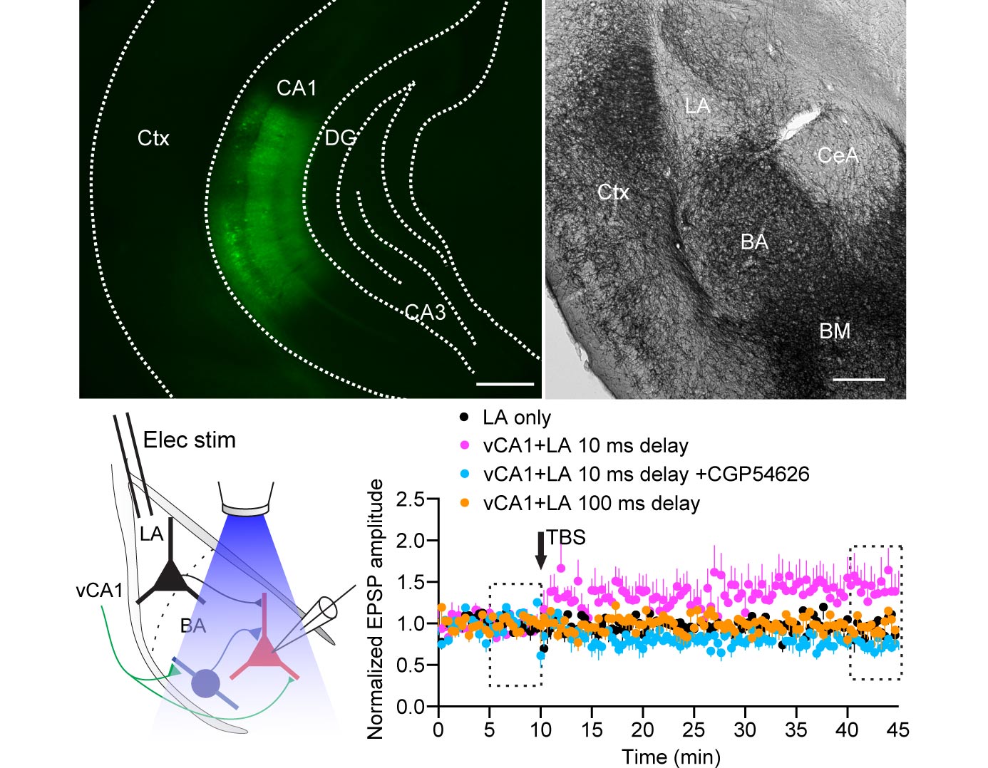
The dynamic interactions between hippocampus and amygdala are critical for emotional memory. Theta synchrony between these structures occurs during fear memory retrieval and may facilitate synaptic plasticity, but the cellular mechanisms are unknown. We report that interneurons of the mouse basal amygdala are activated during theta network activity or optogenetic stimulation of ventral CA1 pyramidal cell axons, whereas principal neurons are inhibited. Interneurons provide feedforward inhibition that transiently hyperpolarizes principal neurons. However, synaptic inhibition attenuates during theta frequency stimulation of ventral CA1 fibers, and this broadens excitatory postsynaptic potentials. These effects are mediated by GABAB receptors and change in the Cl(-) driving force. Pairing theta frequency stimulation of ventral CA1 fibers with coincident stimuli of the lateral amygdala induces long-term potentiation of lateral-basal amygdala excitatory synapses. Hence, feedforward inhibition, known to enforce temporal fidelity of excitatory inputs, dominates hippocampus-amygdala interactions to gate heterosynaptic plasticity. VIDEO ABSTRACT.

Hippocampal Theta Input to the Amygdala Shapes Feedforward Inhibition to Gate Heterosynaptic Plasticity.
Theta burst optical stimulation of hippocampal Inputs gates long-term potentiation of excitatory synapses in the amygdala (Top left) Expression of ChR2/YFP in ventral/intermediate CA1. (Top right) Light micrograph showing channelrhodopsin (ChR2)/YFP-positive axons in the amygdaloid complex. Basal amygdala (BA) and basomedial (BM) nuclei showed densest innervation, whereas only few fibers were detectable in the lateral amygdala (LA) and central amygdala (CeA). (Bottom left) Schematic of the experimental configuration: optical stimulation of vCA1 axons in the BA was paired with electrical stimulation of the LA while recording from principal neurons (PNs). (Bottom right) Pairing of LA-vCA1 theta burst stimulation (TBS) induced long-term potentiation (LTP), which did not occur with TBS of the LA only, but also in the presence of CGP54626 or when TBS of the LA was evoked 100 ms after TBS of the vCA1 axons.
Citation
2015.Neuron, 87(6):1290-1303.
Related Content
Publication
Bocchio M, Fucsina G, Oikonomidis L, McHugh SB, Bannerman DM, Sharp T, Capogna M
2015.Neuropsychopharmacology, 40(13):3015-26.
Publication
Blaesse P, Goedecke L, Bazelot M, Capogna M, Pape HC, Jüngling K
2015.J. Neurosci., 35(19):7317-25.
Publication
Nakamura KC, Sharott A, Tanaka T, Magill PJ

2021. J Neurosci, 41(50):10382-10404.

Publication
Garas FN, Shah RS, Kormann E, Doig NM, Vinciati F, Nakamura KC, Dorst MC, Smith Y, Magill PJ, Sharott A
2016. eLife, 5:e16088.