Functional Connectivity of the Pedunculopontine Nucleus and Surrounding Region in Parkinson's Disease.
The pedunculopontine nucleus, a small area near the bottom of the brain, is thought to be important for the control of walking. It has also been a target for therapeutic ‘deep brain stimulation’ in patients who have difficulty with walking and balance. Here, we show how the pedunculopontine nucleus is connected with other strategic brain sites that might influence walking.
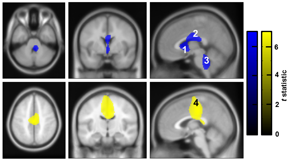
Deep brain stimulation of the pedunculopontine nucleus and surrounding region (PPNR) is a novel treatment strategy for gait freezing in Parkinson's disease (PD). However, clinical results have been variable, in part because of the paucity of functional information that might help guide selection of the optimal surgical target. In this study, we use simultaneous magnetoencephalography and local field recordings from the PPNR in seven PD patients, to characterize functional connectivity with distant brain areas at rest. The PPNR was preferentially coupled to brainstem and cingulate regions in the alpha frequency (8-12 Hz) band and to the medial motor strip and neighboring areas in the beta (18-33 Hz) band. The distribution of coupling also depended on the vertical distance of the electrode from the pontomesencephalic line: most effects being greatest in the middle PPNR, which may correspond to the caudal pars dissipata of the pedunculopontine nucleus. These observations confirm the crucial position of the PPNR as a functional node between cortical areas such as the cingulate/ medial motor strip and other brainstem nuclei, particularly in the dorsal pons. In particular they suggest a special role for the middle PPNR as this has the greatest functional connectivity with other brain regions.
2017.Cereb. Cortex, 27(1):54-67.
2021. Nat Commun, 12(1):5185.