Brain tissue oxygen amperometry in behaving rats demonstrates functional dissociation of dorsal and ventral hippocampus during spatial processing and anxiety.
Damage to a brain region called the hippocampus impairs spatial memory and also reduces anxiety. In this study, we simultaneously recorded activity from two different areas of the hippocampus, and found that the dorsal region was more active during spatial processing whereas the ventral region was more active during anxiety. We conclude there is a regional segregation of hippocampal function.
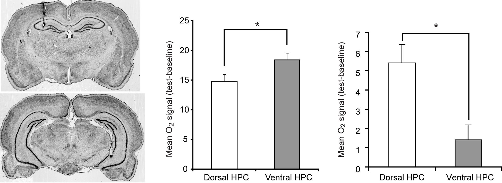
Traditionally, the function of the hippocampus (HPC) has been viewed in unitary terms, but there is growing evidence that the HPC is functionally differentiated along its septotemporal axis. Lesion studies in rodents and functional brain imaging in humans suggest a preferential role for the septal HPC in spatial learning and a preferential role for the temporal HPC in anxiety. To better enable cross-species comparison, we present an in vivo amperometric technique that measures changes in brain tissue oxygen at high temporal resolution in freely-moving rats. We recorded simultaneously from the dorsal (septal; dHPC) and ventral (temporal; vHPC) HPC during two anxiety tasks and two spatial tasks on the radial maze. We found a double-dissociation of function in the HPC, with increased vHPC signals during anxiety and increased dHPC signals during spatial processing. In addition, dHPC signals were modulated by spatial memory demands. These results add a new dimension to the growing consensus for a differentiation of HPC function, and highlight tissue oxygen amperometry as a valuable tool to aid translation between animal and human research.
2011.Eur. J. Neurosci., 33(2):322-37.
2025. Cell Rep, 44(6):115808.

2024. J Neurol Neurosurg Psychiatry, 95(12):1112-1122.