Event-related functional magnetic resonance spectroscopy.
Proton-Magnetic Resonance Spectroscopy (MRS) measures brain chemicals in the human brain. Traditionally, MRS has been used to measure slow changes in brain chemicals, but functional MRS can now be used to measure more rapid changes. This paper provides a user guide for measuring rapid changes in brain chemicals, including those that accompany computations relevant for human cognition and behaviour.
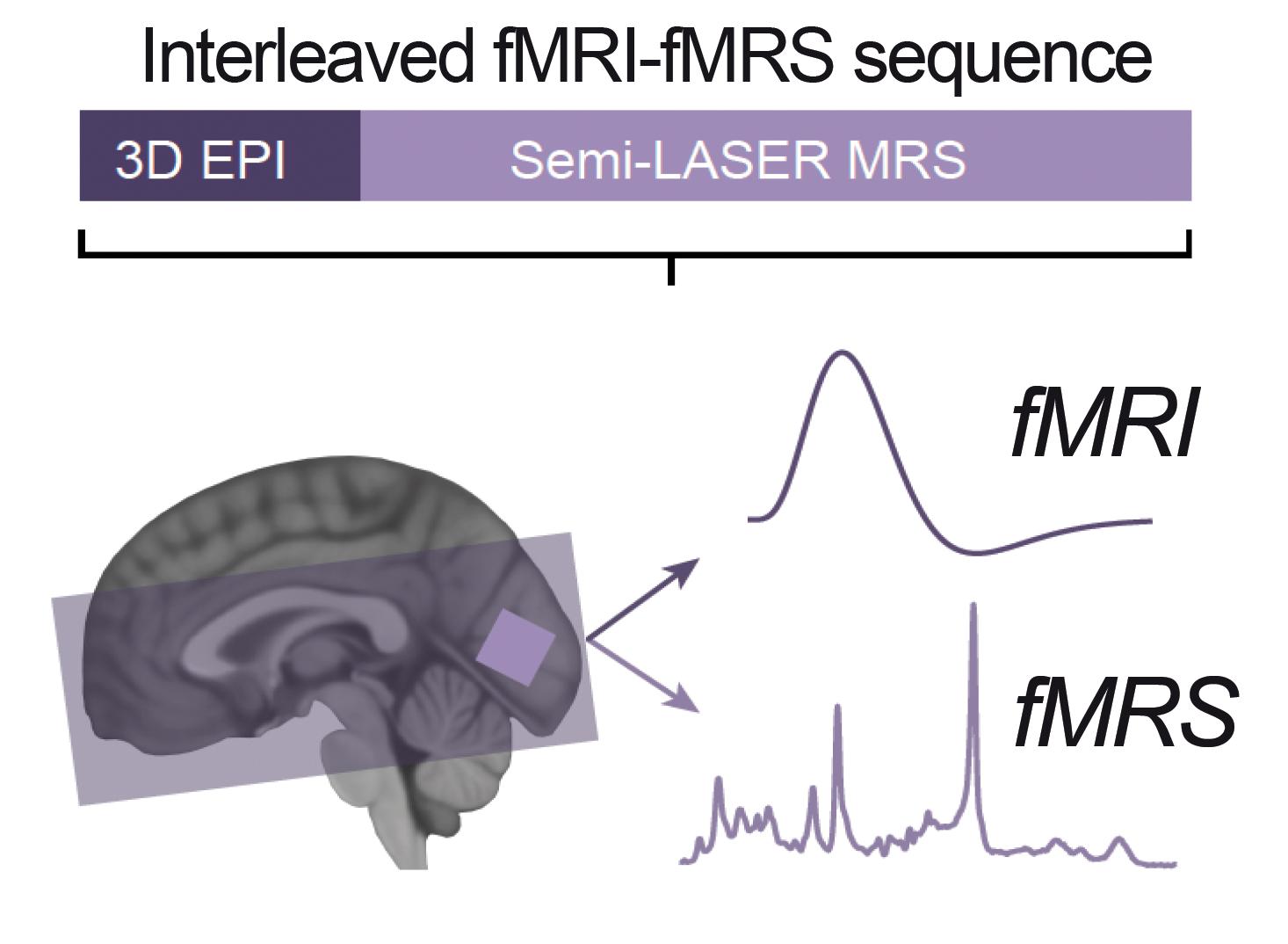
Proton-Magnetic Resonance Spectroscopy (MRS) is a non-invasive brain imaging technique used to measure the concentration of different neurochemicals. "Single-voxel" MRS data is typically acquired across several minutes, before individual transients are averaged through time to give a measurement of neurochemical concentrations. However, this approach is not sensitive to more rapid temporal dynamics of neurochemicals, including those that reflect functional changes in neural computation relevant to perception, cognition, motor control and ultimately behaviour. In this review we discuss recent advances in functional MRS (fMRS) that now allow us to obtain event-related measures of neurochemicals. Event-related fMRS involves presenting different experimental conditions as a series of trials that are intermixed. Critically, this approach allows spectra to be acquired at a time resolution in the order of seconds. Here we provide a comprehensive user guide for event-related task designs, choice of MRS sequence, analysis pipelines, and appropriate interpretation of event-related fMRS data. We raise various technical considerations by examining protocols used to quantify dynamic changes in GABA, the primary inhibitory neurotransmitter in the brain. Overall, we propose that although more data is needed, event-related fMRS can be used to measure dynamic changes in neurochemicals at a temporal resolution relevant to computations that support human cognition and behaviour.
2023. Neuroimage, 276:120194.