When do bursts matter in the primary motor cortex? Investigating changes in the intermittencies of beta rhythms associated with movement states.
The brain's rhythmic electrical activity changes when we prepare, execute, or imagine a movement. We found that short bursts of activity in the motor cortex can tell us about these changes. Using a computer model, we also found that these bursts relate to how the brain balances between internal activity and incoming sensory information.
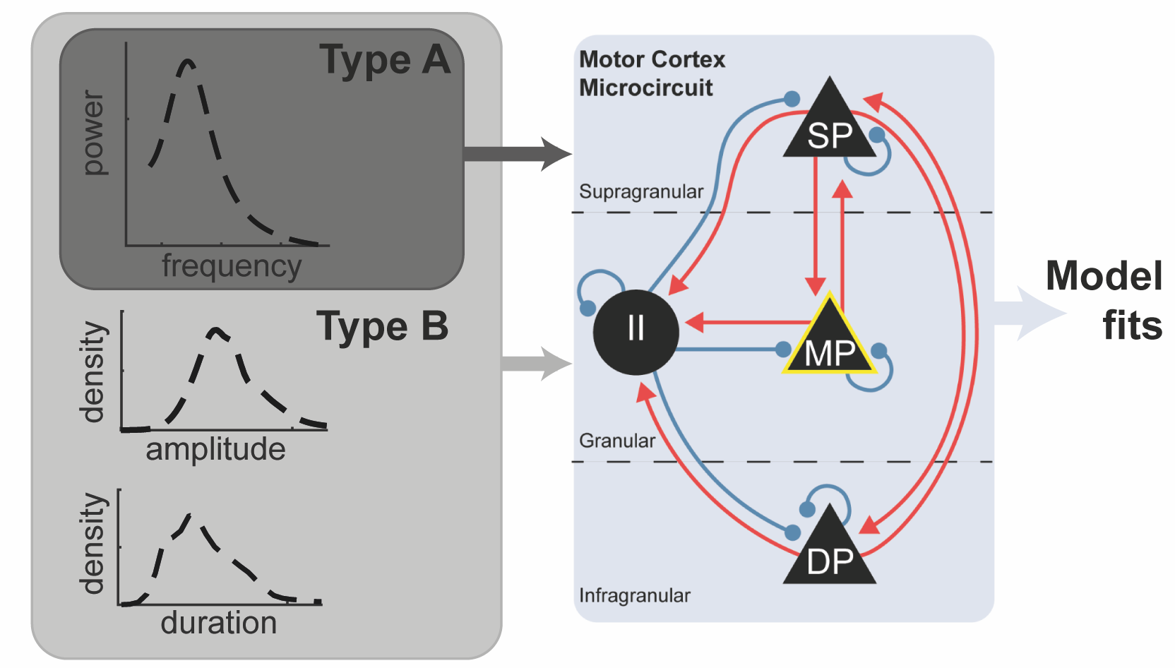
Brain activity exhibits significant temporal structure that is not well captured in the power spectrum. Recently, attention has shifted to characterising the properties of intermittencies in rhythmic neural activity (i.e. bursts), yet the mechanisms that regulate them are unknown. Here, we present evidence from electrocorticography recordings made over the motor cortex to show that the statistics of bursts, such as duration or amplitude, in the beta frequency (14-30 Hz) band, significantly aid the classification of motor states such as rest, movement preparation, execution, and imagery. These features reflect nonlinearities not detectable in the power spectrum, with states increasing in nonlinearity from movement execution to preparation to rest. Further, we show using a computational model of the cortical microcircuit, constrained to account for burst features, that modulations of laminar specific inhibitory interneurons are responsible for the temporal organisation of activity. Finally, we show that the temporal characteristics of spontaneous activity can be used to infer the balance of cortical integration between incoming sensory information and endogenous activity. Critically, we contribute to the understanding of how transient brain rhythms may underwrite cortical processing, which in turn, could inform novel approaches for brain state classification, and modulation with novel brain-computer interfaces.
2023. Prog Neurobiol, 221:102397.
2016.Cereb. Cortex, 26(10):3977-90.
2017.Exp Brain Res, 235(5):1455-1465.